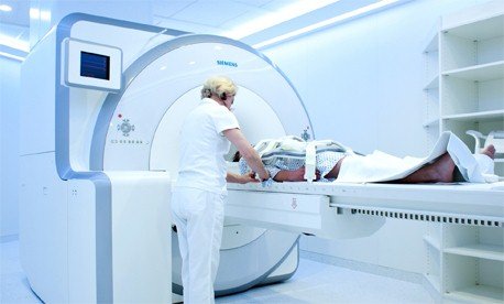
The combination of a magnetic resonance tomography (MR) and a positron emission tomography (PET) in one device allows doctors – for the first time – to simultaneously see the position of internal organs, how these are working, as well as their metabolism, all in a single image. This may help doctors to make a more accurate diagnoses by not only seeing where a tumor is in the body, but also its type and its activity.
Moreover it may display how the body reacts to medication administered to the patient. The device, called the Biograph mMR, has been developed by Siemens Healthcare and now has been installed at the university hospital Klinikum rechts der Isar in Munich. The Biograph mMR combines two technologies that normally would not be able to work next to each other: Magnetic resonance imaging uses a strong magnetic field and electromagnetic waves, while positron emission tomography uses low-dose radioactively charged radiopharmaceuticals, with which the patient is injected before the examination. These radiopharmaceuticals react with the body tissue and the resulting radiation is measured and finally converted into an image. According to physics applied in these imaging techniques, those two technologies should conflict with each other and make simultaneous imaging impossible. But the Biograph mMR is designed to overcome this physical hurdle.
Thanks to funding of the Deutsche Forschungsgemeinschaft (DFG) the first systems are installed in Germany. The combination of both technologies in one system is expected to change the diagnosis of many diseases, including many types of cancer as well as dementia. The Klinikum rechts der Isar has started clinical use testing in an effort to diagnose diseases at a very early stage; to see the progression of disease and to use that information to develop a therapy plan precisely focused on the respective patient. The radiologists plan to use the system for cancer follow-up in the long run, by reducing radiation exposure by the use of the system. In addition, the combination of the two systems cuts down the time needed for an examination nearly by half compared to when two separate systems are used. The same works for the space – where room was needed for two large machines before, now only one combined machine is required. That gives hospitals more space for patients.
This may be a good place to introduce yourself and your site or include some credits.
Search
Automation Training
Our foundation-to-advanced automation course training covers end-to-end industrial workflows used in modern plants. Learners practice the full cycle from basic circuits to commissioning and maintenance with hands-on labs, project-based fault finding, SOP creation, and documentation exposure (URS, FDS, FAT/SAT).
PLC Training
This PLC training builds controller fundamentals with ladder, FBD, and ST, including I/O wiring, PID tuning, diagnostics, and version control practices on live rigs.
SCADA Training
Our SCADA course covers tag databases, HMI graphics, historian/trends, alarm rationalization, redundancy, user security, backups, and deployment aligned to plant standards.
Panel Designing
This panel design course teaches standards-compliant MCC/PLC panel engineering, SLD/GA/wiring docs, device selection, heat-load, testing, and FAT.
BMS & Security
BMS training focuses on HVAC/lighting/utilities automation; CCTV & security covers design, storage, networking, and analytics.
IIoT
The Industrial IoT diploma spans sensors-to-dashboard pipelines: MQTT/OPC UA, gateways, historians, alerts/KPIs, and predictive maintenance basics.